
In this article “Neck Anatomy: The Complete Guide” we will discuss neck anatomy in detail. Read this article to learn lateral cervical region, anterior cervical region, and posterior cervical region. This article includes the following:
Introduction to the Neck
The neck is a vital part of the human body. It’s like a bridge between our head and the rest of our body. It allows us to move our → heads, breathe, eat, and even speak. This vital area comprises → bones, muscles, and other structures.
In the next part, we will learn about the lateral cervical side, which includes → various muscles, important blood vessels, and nerves.
Lateral Cervical Region
The lateral cervical region is like a side area on your neck. It’s important because it’s home to various → muscles, blood vessels, and nerves. Together they play significant roles in how our neck functions. Let’s break down its → boundaries, muscles, blood vessels, and nerves, in a simple way:

Boundaries of the Lateral Cervical Region
This region is defined by the sternocleidomastoid muscle anteriorly and the trapezius muscle posteriorly. It extends from the base of the skull (mastoid process) to the clavicle. Let’s understand both boundaries:
- Anterior Boundary: The sternocleidomastoid muscle (SCM) plays a pivotal role in defining the anterior boundary of this region. The SCM is a large, superficial muscle that extends diagonally across the neck. Then originates from the sternum (breastbone) and clavicle (connects the sternum to the shoulder). And at last, gets inserted in the mastoid process (serves as an attachment for neck muscles) of the temporal bone (cranial bone houses the ear structures). It effectively divides the neck into anterior and posterior triangles.
- Posterior Boundary: The trapezius muscle constitutes the posterior boundary of the lateral cervical region. It is a large, triangular-shaped muscle that covers the upper back and neck. It starts from the occipital bone (back of the skull) and the cervical (neck) or thoracic (vertebral column) vertebrae. And then inserts into the clavicle and scapula.
Muscles of the Lateral Neck
Several muscles call the lateral cervical region home. These muscles help us → move our heads, nod, and even shrug our shoulders. The following are the muscles:

- Sternocleidomastoid Muscle – It’s the muscle that pops out when we turn our head to the side. It helps tilt and rotate our heads.
- Trapezius Muscle– It forms a triangle shape on our upper back. It helps us → shrug, move our shoulders, and maintain good posture.
- Scalene Muscles– The scalene muscles consist of three parts → anterior, middle, and posterior. They lie deep within the lateral cervical region. And they are involved in elevating the ribs during breathing and assisting in neck flexion and rotation.

Important Blood Vessels
The lateral cervical region accommodates vital blood vessels. The external carotid artery supplies blood to the → face, scalp, and neck. Meanwhile, the jugular vein assists in draining blood from the → brain and face. Two significant blood vessels in this region are:
- External Carotid Artery (ECA)– The ECA is a major artery that provides blood supply to the → neck, face, and scalp. It includes → superior thyroid artery (thyroid gland), lingual artery (tongue), facial artery (face), and occipital artery (back of the scalp and neck).
- Internal Carotid Artery (ICA)– ICA does not traverse the lateral cervical side directly. It enters the skull to supply blood to the brain and eyes. It is essential to mention the ICA as it runs closely with the lateral cervical region’s structures.
Nerves in the Lateral Neck
Several nerves traverse the lateral cervical region, serving diverse functions. The accessory nerve innervates the sternocleidomastoid and trapezius muscles, contributing to their movement. The cervical plexus distributes sensory and motor nerves to the skin and muscles of the neck. Several important nerves innervate the lateral cervical region, and those are:
- Accessory Nerve (Cranial Nerve XI)– The accessory nerve primarily innervates the trapezius and sternocleidomastoid muscles. It enables movements like head rotation and shoulder shrugging.
- Cervical Plexus– The cervical plexus is a network of nerves formed by the anterior rami of the upper four cervical spinal nerves (C1-C4). It supplies the skin and muscles of the neck, and its branches, such as the → lesser occipital nerve and supraclavicular nerves. They are essential in providing sensory information from the lateral cervical region.
In the upcoming section, we will learn about the anterior cervical side, which includes → boundaries, various muscles, the thyroid gland, important blood vessels, and nerves.
Anterior Cervical Region
The anterior cervical region is vital at the front of the neck. It encompasses several key anatomical structures, including → muscles, glands, blood vessels, and nerves. Each of them plays a crucial role in supporting various bodily functions. Let’s talk about the various components of this region:
Boundaries of the Anterior Cervical Region
This region extends from the bottom of the jawbone (mandible) to the top of the collarbone (clavicle). It is bordered laterally by the sternocleidomastoid muscles (SCM). While SCM runs diagonally from behind the ear to the collarbone. This region also encompasses the midline of the neck, where the thyroid gland is located.
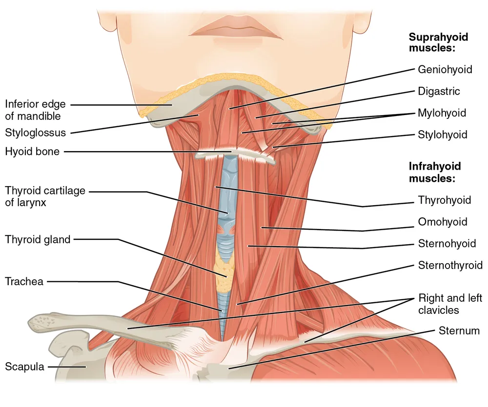
Muscles of the Anterior Neck
The anterior cervical region is home to several muscles that aid in → swallowing, speech, and neck movements. The prominent muscles of the anterior neck are:
- Sternocleidomastoid Muscle: This muscle is one of the most prominent muscles in the anterior neck. It runs diagonally from the sternum and clavicle to the mastoid process of the temporal bone. Each side of the neck has its own sternocleidomastoid muscle. These muscles work together to flex the neck forward, rotate it to the opposite side, and tilt it laterally.
- Thyroid Gland Muscles: The thyroid gland is situated in the front of the neck and is surrounded by some muscles that provide support. These muscles include the infrahyoid muscles (sternohyoid, omohyoid, thyrohyoid, and sternothyroid). They help depress the hyoid bone and stabilize the thyroid cartilage.
- Hyoid Muscles: The hyoid bone is a U-shaped bone located at the base of the tongue and above the thyroid cartilage. Muscles that attach to the hyoid bone, such as the mylohyoid and geniohyoid. They are involved in actions like swallowing and controlling the position of the tongue.
- Digastric Muscle: The digastric muscle consists of two bellies (anterior and posterior) connected by an intermediate tendon. It attaches to the mandible and the hyoid bone. This muscle helps open the mouth, depress the mandible, and stabilize the hyoid bone during swallowing.
- Sternohyoid Muscle: This muscle lies deep in the sternocleidomastoid. It helps in depressing the hyoid bone and larynx during swallowing.
- Sternothyroid Muscle: It is located deeper than the sternohyoid. This muscle assists in depressing the larynx during swallowing and speaking.
- Omohyoid Muscle: The omohyoid muscle consists of superior and inferior bellies. These bellies are separated by an intermediate tendon. It assists in depressing the hyoid bone and larynx, aiding in swallowing.
- Mylohyoid Muscle: Forming the floor of the mouth, the mylohyoid muscle extends from the mylohyoid line of the mandible to the hyoid bone. It aids in swallowing and elevates the floor of the mouth.
- Thyrohyoid Muscle: Connecting the thyroid cartilage to the hyoid bone, the thyrohyoid muscle raises the larynx during swallowing and speech.
- Platysma Muscle: This thin, flat muscle covers the front of the neck and extends to the lower face. It helps in expressions such as frowning and tension of the neck skin.
- Geniohyoid Muscle: Positioned just above the mylohyoid muscle, the geniohyoid muscle connects the mandible to the hyoid bone. It aids in depressing and stabilizing the hyoid bone during swallowing and opening the mouth.
- Stylohyoid Muscle: The stylohyoid muscle is a slender muscle. It originates from the styloid process of the temporal bone (a bony projection located near the ear). It extends downward and attaches to the body of the hyoid bone. The stylohyoid muscle is involved in elevating the hyoid bone during swallowing and speech. It also assists in maintaining the stability of the hyoid bone and the floor of the mouth.

The Thyroid Gland and Its Significance
It is located in the midline of the anterior cervical region, just below the thyroid cartilage. This gland plays a crucial role in → regulating metabolism and energy production in the body. It produces hormones (T3 and T4) that → influence growth, development, and the body’s overall metabolic rate. Thyroid function affects everything from heart rate to body temperature, thus helping in maintaining bodily balance.

Essential Blood Vessels and Nerves
The anterior cervical region contains important blood vessels and nerves. The carotid arteries are located on each side of the neck. They supply oxygen-rich blood to the brain. The jugular veins run parallel to the carotid arteries. They aid in the removal of deoxygenated blood. Nerves in this region include the vagus nerve, which controls various vital functions such as → heart rate and digestion.
In the next section, we will discuss the posterior cervical region, which includes the → boundaries, various muscles, occipital triangle, blood supply, and nerve innervation.
Posterior Cervical Region
The posterior cervical region is a vital part of our neck’s anatomy. It encompasses various structures, including → muscles, blood vessels, and nerves. Let’s delve into its components:
Boundaries of the Posterior Cervical Region
The posterior cervical region is located at the back of the neck. It extends from the base of the skull (occipital bone) to the upper part of the thoracic spine. It is bordered by the trapezius muscle on the sides and the midline of the neck’s vertebrae at the back.

Muscles of the Posterior Neck
Several important muscles are found in the posterior cervical region. These muscles help in major shoulder and neck movements. The following are the muscles in this region:
- Trapezius Muscle– A large, triangular muscle that spans the posterior cervical region and extends down the thoracic spine. The thoracic spine is the middle section of the vertebral column. It conducts movements in the shoulder like → elevation, depression, retraction, and rotation of the scapula.
- Levator Scapulae– The levator scapulae muscle lies deeper into the trapezius. Then runs from the upper cervical vertebrae to the medial border of the scapula. It aids in elevating and adducting the scapula, helping to stabilize the shoulder.
- Splenius Muscles– The splenius muscles consist of the splenius capitis and splenius cervicis. These muscles are situated along the back of the neck and head. They play a crucial role in → neck extension, lateral flexion, and rotation.
Occipital Triangle and Its Contents
The occipital triangle is a diamond-shaped area formed by various muscles. Those muscles are → trapezius muscle, the sternocleidomastoid muscle, and the omohyoid muscle. Inside this triangle, you can find important structures like:
- Accessory Nerve (Cranial Nerve XI): The accessory nerve innervates the sternocleidomastoid and trapezius muscles. And it plays a vital role in head and shoulder movement.
- Occipital Artery: It is a branch of the external carotid artery. It supplies blood to the posterior scalp and muscles in the occipital triangle.
Blood Supply and Nerve Innervation
Blood vessels provide the posterior cervical region with the oxygen and nutrients it needs. Branches of arteries like the → occipital artery and the superficial cervical artery nourish this area. These vessels ensure that muscles and nerves receive proper nourishment to function effectively.
The lesser and the greater occipital nerve provide sensory information. Generally, from the skin on the back of the head and upper neck. These nerves help us feel sensations like → touch, temperature, and pain.
Without knowing we have reached the conclusion. So, let’s recap everything in brief.
Conclusion
The neck is a critical region of the human body that connects the head to the torso and houses numerous vital structures. The neck anatomy include the → lateral cervical region, anterior cervical region, and posterior cervical region. These regions help medical professionals to perform accurate clinical examinations and procedures.
The lateral cervical region is bordered by the → sternocleidomastoid muscle (SCM) (anterior) and the trapezius muscle (posterior). The lateral cervical region is innervated by the accessory nerve (Cranial Nerve XI) and the cervical plexus.
The anterior cervical region is located at the front of the neck. And it is bounded by the → mandible, the anterior surface of the neck, and the sternocleidomastoid muscle on both sides. Key muscles in this area are the infrahyoid and suprahyoid muscles.
The thyroid gland helps in regulating metabolism through thyroid hormones. The common carotid arteries and external jugular veins are significant blood vessels in this region. Branches of the cervical plexus and cervical sympathetic trunk provide nerve innervation.
The posterior cervical region extends from the base of the skull to the first thoracic vertebra. But it is laterally limited by the trapezius muscle. There are various muscles in this region, including the → trapezius, levator scapulae, and splenius muscles. The occipital triangle, located in this area, contains structures like the accessory nerve and the occipital artery.
Blood supply to the posterior cervical region is provided by branches of the external carotid artery. While nerve innervation includes → the greater occipital nerve and dorsal rami of cervical spinal nerves.
With this guide, medical professionals can give optimal care and ensure the well-being of their patients.
Further Reading
We express our heartfelt gratitude to our readers for their unwavering support in engaging with the Intake Learn article on Anatomy. We will continuously provide significant information you can check articles like and .
For more information on this topic, you can check other sources:
- Wikipedia: https://en.wikipedia.org/wiki/_Neck
- Wikipedia: https://en.wikipedia.org/wiki/Human_Anatomy
- Wikipedia: https://en.wikipedia.org/wiki/Human_anatomical_terms
- Wikipedia: https://en.wikipedia.org/wiki/Human_anatomical_parts_named_after_people
- Wikipedia: https://en.wikipedia.org/wiki/Head_and_neck_anatomy
Attribution
- OpenStax College, CC BY 3.0, via Wikimedia Commons
- Mikael Häggström.When using this image in external works, it may be cited as:Häggström, Mikael (2014). “Medical gallery of Mikael Häggström 2014”. WikiJournal of Medicine 1 (2). DOI:10.15347/wjm/2014.008. ISSN 2002-4436. Public Domain.orBy Mikael Häggström, used with permission., Public domain, via Wikimedia Commons
- OpenStax, CC BY 4.0, via Wikimedia Commons
- Henry Vandyke Carter, Public domain, via Wikimedia Commons
- OpenStax College, CC BY 3.0, via Wikimedia Commons
0 Comments